The Brain
Three cavities, called the primary brain vesicles, form during the early embryonic development of the brain. These are the forebrain (prosencephalon), the midbrain (mesencephalon), and the hindbrain (rhombencephalon).
During subsequent development, the three primary brain vesicles develop into five secondary brain vesicles. The names of these vesicles and the major adult structures that develop from the vesicles follow (see Table 1 ):
-
The telencephalon generates the cerebrum (which contains the cerebral cortex, white matter, and basal ganglia).
-
The diencephalon generates the thalamus, hypothalamus, and pineal gland.
-
The mesencephalon generates the midbrain portion of the brain stem.
-
The metencephalon generates the pons portion of the brain stem and the cerebellum.
-
The myelencephalon generates the medulla oblongata portion of the brain stem
.TABLE 1 The Vesicles and Their Components Primary Vesicles
Secondary Vesicles
Adult Structure
Important Components or Features
prosencephalon (forebrain)
telencephacerebrum
cerebral (cerebral hemispheres)
cerebral cortex (gray matter): motor areas, sensory areas, association areas
prosencephalon (forebrain)
telencephacerebrum
cerebral (cerebral hemispheres)
cerebral white matter: association fibers, commisural fibers, projection fibers
prosencephalon (forebrain)
telencephacerebrum
cerebral (cerebral hemispheres)
basal ganglia (gray matter): caudate nucleus & amygdala, putamen, globus pallidus
prosencephalon
diencephalon
diencephalon
thalamus: relays sensory information
prosencephalon (forebrain)
diencephalon
diencephalon
hypothalamus: maintains body homeostasis
prosencephalon (forebrain)
diencephalon
diencephalon
mammillary bodies: relays sensations of smells to cerebrum
prosencephalon (forebrain)
diencephalon
diencephalon
optic chiasma: crossover of optic nerves
prosencephalon (forebrain)
diencephalon
diencephalon
infundibulum: stalk of pituitary gland
prosencephalon (forebrain)
diencephalon
diencephalon
pituitary gland: source of hormones
prosencephalon (forebrain)
diencephalon
diencephalon
epithalamus: pineal gland
mesencephalon (midbrain)
mesencephalon
brain stem
midbrain: cerebral peduncles, sup. cerebellar peduncles, corpora quadrigemina, superior colliculi
rhombencephalon (hindbrain)
metencephalon
brain stem
pons: middle cerebellar peduncles, pneumotaxic area, apneustic area
rhombencephalon (hindbrain)
metencephalon
cerebellum
sup. cerebellar peduncles, middle cerebellar peduncles, inferior cerebellar peduncles
rhombencephalon (hindbrain)
myelencephalon
brain stem
medulla oblongata: pyramids, cardiovascular center, respiratory center
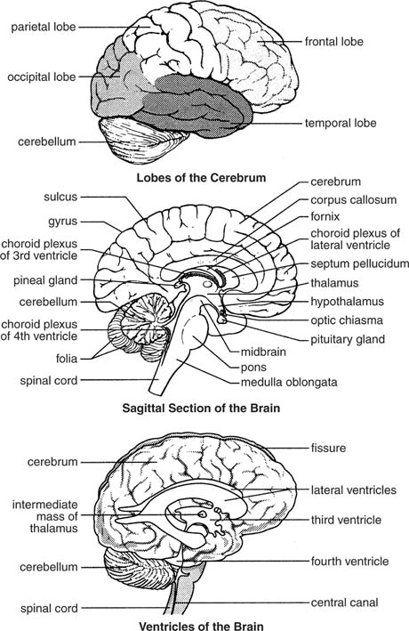
A second method for classifying brain regions is by their organization in the adult brain. The following four divisions are recognized (see Figure 1 ).
| | |||
| |||
| | |||
| |||
| |
-
-
A gyrus (plural, gyri) is an elevated ridge among the convolutions.
-
A sulcus (plural, sulci) is a shallow groove among the convolutions.
-
A fissure is a deep groove among the convolutions.
A cross section of the cerebrum shows three distinct layers of nervous tissue:
-
-
The following structures are either included or associated with the hypothalamus.
-
The mammillary bodies relay sensations of smell.
-
The infundibulum connects the pituitary gland to the hypothalamus.
-
-
The brain stem connects the diencephalon to the spinal cord. The brain stem resembles the spinal cord in that both consist of white matter fiber tracts surrounding a core of gray matter. The brain stem consists of the following four regions, all of which provide connections between various parts of the brain and between the brain and the spinal cord. (Some prominent structures are illustrated in Figure 2 ).

Figure 2 Prominent structures of the brain stem.
-
The midbrain is the uppermost part of the brain stem.
-
The pons is the bulging region in the middle of the brain stem.
-


0 comments:
Post a Comment